


An ultrasound scan, sometimes called a sonogram, is a procedure that uses high-frequency sound waves to create an image of part of the inside of the body. An ultrasound scan can be used to monitor an unborn baby, diagnose a condition, or guide a surgeon during certain procedures. An ultrasound scan uses high-frequency sound waves to create images of the inside of the body. It is suitable for use during pregnancy.
Ultrasound scans, or sonography, are safe because they use sound waves or echoes to make an image, instead of radiation. Ultrasound scans are used to evaluate fetal development, and they can detect problems in the liver, heart, kidney, or abdomen. They may also assist in performing certain types of biopsy. The image produced is called a sonogram.
Fast facts on ultrasound scans:
- Ultrasound scans are safe and widely used.
- They are often used to check the progress of a pregnancy.
- They are used for diagnosis or treatment.
- No special preparation is normally necessary before an ultrasound scan.
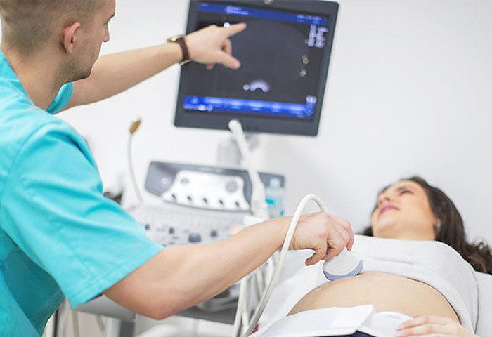
- The person who performs an ultrasound scan is called a sonographer, but the images are interpreted by radiologists, cardiologists, or other specialists.
- The sonographer usually holds a transducer, a hand-held device, like a wand, which is placed on the patient’s skin.
- Ultrasound is sound that travels through soft tissue and fluids, but it bounces back, or echoes, off denser surfaces. This is how it creates an image.
- The term “ultrasound” refers to sound with a frequency that humans cannot hear.
- For diagnostic uses, the ultrasound is usually between 2 and 18 megahertzTrusted Source (MHz).
- Higher frequencies provide better quality images but are more readily absorbed by the skin and other tissue, so they cannot penetrate as deeply as lower frequencies
- Lower frequencies penetrate deeper, but the image quality is inferior.
How does it capture an image?
- Ultrasound will travel through blood in the heart chamber, for example, but if it hits a heart valve, it will echo, or bounce back.
- It will travel straight through the gallbladder if there are no gallstones, but if there are stones, it will bounce back from them.
- The denser the object the ultrasound hits, the more of the ultrasound bounces back.
- This bouncing back, or echo, gives the ultrasound image its features. Varying shades of gray reflect different densities.